BrainLoc 6.0
3D Dipole Localization Software
Description
Functions:
- localization of pathological electrical activity sources when suffering from epilepsy, injuries, insults, neoformations
- localization of evoked potentials sources, wave patterns, rhythmic activity generators
Methods of Data Editing:
- selection of the arbitrary fragment of the recorded EEG/EP for the detailed analysis
- removal of the channels with the artifacts
- correction of the electrode names and settings
- capability of the recorded EEG/EP inversion
- isoline correction of the whole file or specified interval (for example, basing on the prestimulative EP interval)
- signal resampling
- signal filtering in the standard ranges or arbitrary frequency band
- calculating of the power spectrum of the whole record (or its part) for the detection of the power spectral components sources
Models Applied for Data Analysis:
- model with non-stationary dipoles
- model with stationary dipoles
Models with Non-stationary Dipoles:
- for every time point of the analyzed data its own dipole model is calculated which includes one or two non-stationary dipoles
- it is possible to specify the averaging window according to time when calculating the dipole sources
Model with Stationary Dipoles:
- single model with the given number of dipoles (from one to eight) for the whole data fragment is calculated;
- model variants:
- dipole centers are linked with the given structures
- search of the optimal arrangement of the dipole centers in the brain volume
- search of the steady optimal dipole orientation for the whole data fragment
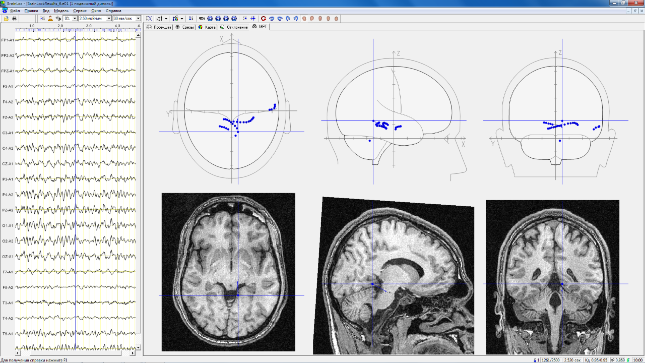
Visualization of Calculated Dipole Sources:
- on three head views
- on diagrammatic sectional views of the brain structures
- on tomographic brain images (MRT-images)
- validity assessment of the calculated parameters and the automatic selection of the reliable sources
- displaying the total information about the dipole model parameters
- mapping of the dipole ratio distribution on the axial sections
- simultaneous review of the several files in the multiwindowing
Amplitude Mapping of:
- EEG and EP on the head surface
- EEG and EP calculated on the cortex surface
- dipole model potentials and rest potentials
- Graphs Displaying of:
- EEG and EP traces
- dipole model potentials and rest potentials
- dipole moment projections on the coordinates and the dipole moment modules
- Data Input:
- EEG and EP data file reading recorded in different binary and ASCII formats, the European data format (EDF) supporting
- МRТ- и CT brain images input, recorded on the different types of tomographes
- Additional Capabilites:
- creation of EEG and EP test files with the given dipole model for the examination of the direct and inverse tasks solving
Hardware Configuration:
IBM PC Pentium (or compatible) with MS Windows 9x/NT/2000/XP operational system
The program got the research results verification in the leading medical research institutes and clinics: N.N. Burdenko Institute of Neurosurgery, Institute of Neurology, I.M. Sechenov Moscow Medical Academy, N.V. Skliphosovsky Ambulance Research Center, Moscow Research Institute of Psychiatry of the Ministry of Health of the Russian Federation, Institute for Neurosurgical Research Named after A.L. Polenov, and etc.